|
시장보고서
상품코드
2124879
공초점 현미경 : 시장 점유율 분석, 업계 동향 및 통계, 성장 예측(2026-2031년)Confocal Microscope - Market Share Analysis, Industry Trends & Statistics, Growth Forecasts (2026 - 2031) |
||||||
 Mordor Intelligence
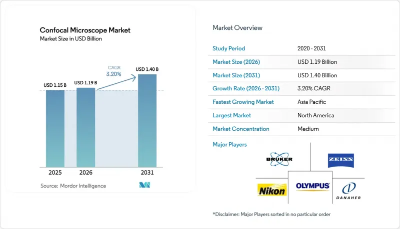
Mordor Intelligence에 의하면, 공초점 현미경 시장 규모는 2025년 11억 5,000만 달러로 평가되었습니다. 2026년 11억 9,000만 달러에서 2031년까지 14억 달러로 확대되고 2026-2031년까지 연평균 복합 성장률(CAGR)은 3.20%를 나타낼 전망입니다.
본 보고서는 현미경 유형(레이저 스캐닝 공초점 현미경, 스피닝 디스크형 공초점 현미경, 기타), 광원(다이오드 레이저, OPSL·파이버 레이저, 기타), 최종 사용자(병원 및 클리닉, 진단 실험실, 기타),지역(북미, 유럽, 아시아태평양, 중동 및 아프리카, 남미)별로 분류되어 있습니다. 시장 예측은 금액(달러) 기준으로 제시되어 있습니다.
세계의 공초점 현미경 시장 동향 및 인사이트
안과 의료 분야에서 공초점 이미징 수요 증가
생체 내 각막 공초점 현미경 검사는 연구 용도에서 표준 진단법으로 전환되고 있으며, 특히 당뇨병성 말초 신경병증이나 감염성 각막염의 조기 발견에서 이러한 경향이 두드러집니다. 미국 안과학회는 2025년판 가이드라인을 개정하여, 무증상 신경병증을 보이는 2형 당뇨병 환자에 대한 각막 신경섬유 밀도의 정량화를 권장했습니다. 이에 따라 병원에서는 하이델베르크 레티나 토모그래프나 니덱의 컴포스캔 모듈 도입이 진행되고 있습니다. 메디케어는 2025년 1월, 각막 공초점 현미경 검사에 CPT 코드 92499를 할당하고, 양안 검사당 285달러를 환급함으로써 미국 클리닉의 경제적 장벽을 제거했습니다. 2025년에는 콘택트렌즈와 관련된 미생물성 각막염 사례가 전년 대비 18% 급증했으나, 실시간 영상 진단을 통해 진단 소요 시간이 48시간에서 20분 이하로 단축되었습니다. 인도의 실명 방지 프로그램에서는 2025-26 회계연도에 12억 루피(1,440만 달러)를 편성하여 2급 도시의 안과 의료 센터에 공초점 시스템을 도입할 예정입니다. 이러한 정책 전환은 여러 지역의 공초점 현미경 시장을 견인하고 있습니다.
생명과학 및 신경생리학 연구개발 예산 확대
미국 국립보건원(NIH)의 예산은 2025년에 513억 달러로 증액되었으며, 공초점 현미경이 기초 기술로서 필수적인 신경과학, 암 생물학, 감염병 연구에 중점을 두고 있습니다. 중국 국가자연과학기금은 신경과학 이미징에 35억 위안(4억 9,000만 달러)을 배정하고, 주요 대학에 다광자 공초점 현미경 설치를 의무화했습니다. ‘Horizon Europe’은 2025년부터 2027년까지 생명과학 인프라에 21억 유로(23억 달러)를 배정하며, 첨단 이미징 코어의 구축을 명확히 요구하고 있습니다. 일본의 ‘Brain/MINDS’ 프로젝트에서는 2광자 공초점 시스템을 이용한 마모셋의 커넥톰 매핑에 82억 엔(5,500만 달러)을 배정했습니다. 지속적인 자금 지원은 공급업체의 연구개발 위험을 완화하고, 적응형 광학 및 AI 자동 초점 분야의 혁신을 촉진함으로써 공초점 현미경 시장의 확대로 이어지고 있습니다.
높은 초기 투자 및 유지비와 긴 투자 회수 기간
공초점 현미경의 가격은 엔트리 레벨 스피닝 디스크형의 5만 달러부터 환경 제어 챔버를 갖춘 다광자 플랫폼의 60만 달러까지 폭이 넓어, 커뮤니티 칼리지나 지방 병원의 예산을 초과합니다. 연간 유지보수 계약 비용은 구매 가격의 10%-12%를 차지하며, 레이저 모듈은 8,000-1만 2,000시간마다 교체가 필요하고, 그 비용은 최대 8만 달러에 달할 전망입니다. 인도에서는 20%, 브라질에서는 최대 18%의 수입 관세가 부과되어, 미국 가격과 비교했을 때 총 비용이 30%-40% 더 비쌉니다. 리스 모델도 등장하고 있지만, 도입률은 총 설치 대수의 5% 이하에 그치고 있습니다. 높은 진입 장벽이 공초점 현미경 시장의 단기적인 성장을 억제하고 있습니다.
부문별 분석
2025년, 레이저 스캐닝 시스템은 공초점 현미경 시장의 59.65% 점유율을 차지했습니다. 이는 형광증백제 선택의 유연성과 지난 30년 동안 발표된 프로토콜과의 호환성에 힘입은 결과입니다. Zeiss LSM 980 및 Leica STELLARIS 8 플랫폼은 400nm에서 800nm까지의 스펙트럼 감지가 가능하며, 초당 420프레임을 기록할 수 있어 뉴런의 칼슘 이미징에 없어서는 안 될 존재가 되었습니다. 요코가와(Yokogawa)의 CSU-W1과 같은 스피닝 디스크 현미경은 광 퇴색을 줄이면서 200-500fps를 실현하여 생세포 배아학 연구에 활용되고 있습니다. 브루커(Bruker)의 Ultima Investigator와 같은 2광자 시스템은 2mm 두께의 산란 조직을 투과하여 뇌 매핑 요건을 충족합니다.
프로그래머블 어레이 현미경은 2031년까지 연평균 성장률(CAGR) 4.32%로 확대될 것으로 예측되며, 공초점 현미경 시장 전체를 상회하는 성장이 예상됩니다. 옥스포드 기기(Oxford Instruments)의 ‘Andor Dragonfly’는 스피닝 디스크의 고속성과 STED의 광독성 없이 가로 해상도 100nm에 육박하는 계산 기반 초해상도 기술을 결합하고 있습니다. 스윕 필드 모드와 라인 스캔 모드는 고성능 스크리닝 분야에서 틈새 시장을 차지하고 있습니다. 니콘의 ‘AX R’ 라인 스캔은 96웰 플레이트의 촬영 시간을 40% 단축합니다. ISO 13485 인증을 취득한 공급업체가 진단 기기 입찰에서 수주하며, 선구자로서의 우위를 공고히 하고 있습니다.
지역별 분석
북미는 2025년 매출의 36.71%를 차지했습니다. 이는 510억 달러가 넘는 NIH의 자금 지원과, 공초점 이미징을 청구 가능한 임상 서비스로 규정하는 메디케어의 환급 제도에 힘입은 결과입니다. 미국의 BRAIN 이니셔티브는 이미징 인프라에 1억 4,000만 달러를 배정하고, 22개 기관에 다광자 공초점 시스템 도입을 의무화했습니다. 캐나다의 NSERC는 이미징 핵심 시설에 4억 5,000만 캐나다 달러(3억 3,000만 달러)를 지원했습니다. 한편, 멕시코는 2,500만 달러 규모의 현대화 프로그램을 시작했으나, 15%의 수입 관세로 인해 진전이 저해되고 있습니다.
아시아태평양은 2031년까지 연평균 성장률(CAGR) 4.17%를 나타낼 것으로 예측되어, 주요 지역 중 가장 높은 성장률을 보이고 있습니다. 중국에서는 35억 위안 규모의 신경과학 보조금을 재원으로 2025년에 320대의 공초점 장비가 도입되었으며, 그중 60%는 멀티포톤 기능을 갖추고 있었습니다. 일본의 ‘Brain/MINDS’ 프로젝트는 2025년에 82억 엔(5,500만 달러)을 투자하여 심부 뇌 영상 촬영용 적응 광학 지원 시스템을 도입했습니다. 인도는 첨단 현미경 기술에 18억 루피(2,160만 달러)를 배정했으나, 20%의 관세와 운영 인력 부족으로 인해 이용률은 60%에 그치고 있습니다. 한국의 기초과학연구원(IBS)은 18곳의 이미징 센터 간 데이터 공유를 효율화하기 위해 자이스(Zeiss)와 라이카(Leica)를 표준 장비로 채택했습니다.
독일의 DFG는 STED 지원 플랫폼을 우선시하여 45대의 공초점 현미경 도입에 자금을 지원했습니다. 영국의 BBSRC는 이미징 인프라에 2,800만 파운드(3,500만 달러)를 투자하여 프랜시스 클릭 연구소 및 주요 대학에 자산을 집중시켰습니다. 프랑스의 CNRS는 거점 간 연수를 원활하게 하기 위해 32개 플랫폼에서 라이카 STELLARIS를 표준화했습니다. 사우디아라비아의 KAUST는 6대의 자이스 LSM 980을 도입했고, 남아프리카공화국의 NRF는 2개 대학의 장비 업그레이드에 자금을 지원하여, 해당 지역에서의 도입이 확대되고 있음을 보여주고 있습니다. 환율 변동과 통관 지연이 계속해서 남미의 성장을 저해하고 있으며, 브라질의 ICMS 세금으로 인해 장비 가격에 최대 18%의 추가 부담이 발생하고 있습니다.
기타 혜택
- 엑셀 형식 시장 예측(ME) 시트
- 3개월간의 애널리스트 지원
자주 묻는 질문
목차
제1장 서론
제2장 조사 방법
제3장 주요 요약
제4장 시장 구도
제5장 시장 규모 및 성장 예측
제6장 경쟁 구도
제7장 시장 기회 및 향후 전망
KTH 26.09.14According to Mordor Intelligence, the confocal microscope market size is projected to expand from USD 1.15 billion in 2025 and USD 1.19 billion in 2026 to USD 1.40 billion by 2031, registering a CAGR of 3.20% between 2026 to 2031.
This report is Segmented by Microscope Type (Laser Scanning Microscopes, Spinning Disk Confocal Microscopes and More), Light Source (Diode Lasers, OPSL & Fiber Lasers, and More) End User (Hospitals & Clinics, Diagnostic Laboratories and More), and Geography (North America, Europe, Asia-Pacific, Middle East and Africa and South America). The Market Forecasts are Provided in Terms of Value (USD).
Global Confocal Microscope Market Trends and Insights
Rising Demand for Ophthalmic-Care Confocal Imaging
In-vivo corneal confocal microscopy is moving from research use toward standard-of-care diagnostics, especially for early detection of diabetic peripheral neuropathy and infectious keratitis. The American Academy of Ophthalmology updated its 2025 guidelines to recommend corneal-nerve fiber density quantification for patients with type 2 diabetes showing subclinical neuropathy, prompting hospitals to purchase Heidelberg Retina Tomograph and Nidek Confoscan modules. Medicare assigned CPT 92499 to corneal confocal microscopy in January 2025, reimbursing USD 285 per bilateral exam and removing a financial barrier for U.S. practices. Contact-lens-related microbial keratitis cases surged 18% year-over-year in 2025, and real-time imaging shortens diagnosis from 48 hours to under 20 minutes. India's blindness-control program earmarked INR 120 crore (USD 14.4 million) in fiscal 2025-26 to equip tier-2 city eye-care centers with confocal systems. These policy shifts bolster the confocal microscopy market across multiple geographies.
Expansion of Life-Science & Neurophysiology R&D Budgets
The U.S. National Institutes of Health budget rose to USD 51.3 billion in 2025, with a focus on neuroscience, cancer biology, and infectious disease, where confocal microscopy is foundational. China's National Natural Science Foundation dedicated CNY 3.5 billion (USD 490 million) to neuroscience imaging, mandating the installation of multiphoton confocal microscopes at leading universities. Horizon Europe earmarked EUR 2.1 billion (USD 2.3 billion) for 2025-2027 life-science infrastructure, explicitly calling for advanced imaging cores. Japan's Brain/MINDS project allocated JPY 8.2 billion (USD 55 million) to marmoset connectome mapping using two-photon confocal systems. Sustained funding de-risks vendor R&D, encouraging innovation in adaptive optics and AI autofocus that expands the confocal microscopy market.
High Capital and Maintenance Cost & Long ROI Cycles
Confocal microscopes range from USD 50,000 for entry-level spinning-disk units to USD 600,000 for multiphoton platforms with environmental chambers, exceeding budgets of community colleges and regional hospitals. Annual service contracts consume 10%-12% of the purchase price, and laser modules require replacement every 8,000-12,000 hours at up to USD 80,000. Import duties add 20% in India and up to 18% in Brazil, inflating landed costs by 30%-40% versus U.S. pricing. Leasing models are emerging, yet adoption remains below 5% of total placements. High entry barriers temper near-term growth in the confocal microscopy market.
Other drivers and restraints analyzed in the detailed report include:
- Growing Incidence of Microbial Keratitis & Corneal Disorders
- AI-Enabled Automated Image-Analysis Workflows
- Shortage of Advanced Microscopy Specialists
For complete list of drivers and restraints, kindly check the Table Of Contents.
Segment Analysis
Laser scanning systems captured 59.65% of the confocal microscopy market share in 2025, sustained by flexibility in fluorophore selection and compatibility with three decades of published protocols. Zeiss LSM 980 and Leica STELLARIS 8 platforms offer spectral detection from 400 nm to 800 nm and record 420 frames per second, making them indispensable for calcium imaging in neurons. Spinning-disk microscopes such as Yokogawa CSU-W1 achieve 200-500 fps with reduced photobleaching, serving live-cell embryology studies. Two-photon systems like Bruker Ultima Investigator penetrate 2 mm of scattering tissue, fulfilling brain-mapping requirements.
Programmable array microscopes are forecast to expand at a 4.32% CAGR through 2031, outpacing the overall confocal microscopy market. Oxford Instruments' Andor Dragonfly combines spinning-disk speed with computational super-resolution that approaches 100 nm lateral resolution without the phototoxicity of STED. Swept-field and line-scanning modes occupy niche positions in high-throughput screening; Nikon AX R line scanning reduces 96-well plate acquisition time by 40%. Vendors holding ISO 13485 certification are winning diagnostic tenders, reinforcing first-mover advantages.
Complete Report Scope:
- By Microscope Type
- Laser Scanning Confocal Microscopes
- Spinning-Disk Confocal Microscopes
- Programmable Array Microscopes (PAM)
- Two-Photon / Multiphoton Confocal Microscopes
- Swept-Field & Line-Scanning Confocal Microscopes
- By Light Source
- Diode Lasers
- OPSL & Fiber Lasers
- LED Illumination
- Others
- By End User
- Hospitals & Clinics
- Diagnostic Laboratories
- Academic & Research Institutes
- Pharmaceutical & Biotechnology Companies
- Other End Users
- By Geography
- North America
- United States
- Canada
- Mexico
- Europe
- Germany
- United Kingdom
- France
- Italy
- Spain
- Rest of Europe
- Asia-Pacific
- China
- Japan
- India
- South Korea
- Australia
- Rest of Asia-Pacific
- Middle East & Africa
- GCC
- South Africa
- Rest of Middle East & Africa
- South America
- Brazil
- Argentina
- Rest of South America
- North America
Geography Analysis
North America captured 36.71% of 2025 revenue, underpinned by NIH funding exceeding USD 51 billion and Medicare reimbursement that positions confocal imaging as a billable clinical service. The U.S. BRAIN Initiative allocated USD 140 million to imaging infrastructure, mandating multiphoton confocal systems at 22 institutions. Canada's NSERC directed CAD 450 million (USD 330 million) to imaging cores, while Mexico launched a USD 25 million modernization program, albeit hampered by 15% import tariffs.
Asia-Pacific is projected to grow at a 4.17% CAGR through 2031, the fastest among major regions. China installed 320 confocal units in 2025, 60% with multiphoton capability, fueled by CNY 3.5 billion neuroscience grants. Japan's Brain/MINDS project invested JPY 8.2 billion (USD 55 million) in 2025, installing adaptive-optics-enabled systems for deep-brain imaging. India earmarked INR 180 crore (USD 21.6 million) for advanced microscopy, yet 20% duties and limited operator pools restrict utilization to 60%. South Korea's Institute for Basic Science standardized on Zeiss and Leica to streamline data sharing across 18 imaging centers.
Germany's DFG funded 45 confocal acquisitions, prioritizing STED-capable platforms. The U.K. BBSRC invested GBP 28 million (USD 35 million) in imaging infrastructure, concentrating assets at the Francis Crick Institute and leading universities. France's CNRS standardized on Leica STELLARIS across 32 platforms to ease cross-site training. Saudi Arabia's KAUST installed six Zeiss LSM 980 units, while South Africa's NRF funded upgrades at two universities, indicating emerging regional adoption. Currency volatility and customs delays continue to hinder South American growth, with Brazil's ICMS taxes adding up to 18% to instrument prices.
- Agilent Technologies
- Bruker
- Caliber Imaging & Diagnostics
- Confocal.nl
- CrestOptics S.p.A
- Hitachi High-Tech Corp.
- HORIBA
- ISS
- JEOL Ltd.
- Keyence Corporation
- Danaher
- Molecular Devices
- Nikon
- Olympus Corporation (Evident)
- Oxford Instruments (Andor)
- PerkinElmer (Revvity)
- PicoQuant GmbH
- Thermo Fisher Scientific
- Thorlabs
- Yokogawa Electric
- Carl Zeiss
Additional Benefits:
- The market estimate (ME) sheet in Excel format
- 3 months of analyst support
TABLE OF CONTENTS
1 Introduction
- 1.1 Study Assumptions & Market Definition
- 1.2 Scope of the Study
2 Research Methodology
3 Executive Summary
4 Market Landscape
- 4.1 Market Overview
- 4.2 Market Drivers
- 4.2.1 Rising Demand for Ophthalmic-Care Confocal Imaging
- 4.2.2 Expansion Of Life-Science & Neurophysiology R&D Budgets
- 4.2.3 Growing Incidence of Microbial Keratitis & Corneal Disorders
- 4.2.4 AI-Enabled Automated Image-Analysis Workflows
- 4.2.5 Adoption Of Super-Resolution Modules for Live-Cell Studies
- 4.2.6 Government-Funded Brain-Connectome Mega-Projects
- 4.3 Market Restraints
- 4.3.1 High Capital/Maintenance Cost & Long ROI Cycles
- 4.3.2 Shortage Of Advanced Microscopy Specialists
- 4.3.3 Shift Of Organoid Research Toward Light-Sheet Microscopy
- 4.3.4 Cybersecurity Compliance Burden For Networked Instruments
- 4.4 Supply-Chain Analysis
- 4.5 Regulatory Landscape
- 4.6 Technological Outlook
- 4.7 Porter's Five Forces
- 4.7.1 Threat of New Entrants
- 4.7.2 Bargaining Power of Buyers
- 4.7.3 Bargaining Power of Suppliers
- 4.7.4 Threat of Substitutes
- 4.7.5 Intensity of Competitive Rivalry
5 Market Size & Growth Forecasts (Value, USD)
- 5.1 By Microscope Type
- 5.1.1 Laser Scanning Confocal Microscopes
- 5.1.2 Spinning-Disk Confocal Microscopes
- 5.1.3 Programmable Array Microscopes (PAM)
- 5.1.4 Two-Photon / Multiphoton Confocal Microscopes
- 5.1.5 Swept-Field & Line-Scanning Confocal Microscopes
- 5.2 By Light Source
- 5.2.1 Diode Lasers
- 5.2.2 OPSL & Fiber Lasers
- 5.2.3 LED Illumination
- 5.2.4 Others
- 5.3 By End User
- 5.3.1 Hospitals & Clinics
- 5.3.2 Diagnostic Laboratories
- 5.3.3 Academic & Research Institutes
- 5.3.4 Pharmaceutical & Biotechnology Companies
- 5.3.5 Other End Users
- 5.4 By Geography
- 5.4.1 North America
- 5.4.1.1 United States
- 5.4.1.2 Canada
- 5.4.1.3 Mexico
- 5.4.2 Europe
- 5.4.2.1 Germany
- 5.4.2.2 United Kingdom
- 5.4.2.3 France
- 5.4.2.4 Italy
- 5.4.2.5 Spain
- 5.4.2.6 Rest of Europe
- 5.4.3 Asia-Pacific
- 5.4.3.1 China
- 5.4.3.2 Japan
- 5.4.3.3 India
- 5.4.3.4 South Korea
- 5.4.3.5 Australia
- 5.4.3.6 Rest of Asia-Pacific
- 5.4.4 Middle East & Africa
- 5.4.4.1 GCC
- 5.4.4.2 South Africa
- 5.4.4.3 Rest of Middle East & Africa
- 5.4.5 South America
- 5.4.5.1 Brazil
- 5.4.5.2 Argentina
- 5.4.5.3 Rest of South America
- 5.4.1 North America
6 Competitive Landscape
- 6.1 Market Concentration Analysis
- 6.2 Market Share Analysis
- 6.3 Company Profiles (includes Global level Overview, Market level overview, Core Segments, Financials as available, Strategic Information, Market Rank/Share for key companies, Products & Services, and Recent Developments)
- 6.3.1 Agilent Technologies
- 6.3.2 Bruker Corporation
- 6.3.3 Caliber Imaging & Diagnostics
- 6.3.4 Confocal.nl
- 6.3.5 CrestOptics S.p.A
- 6.3.6 Hitachi High-Tech Corp.
- 6.3.7 Horiba Ltd.
- 6.3.8 ISS Inc.
- 6.3.9 JEOL Ltd.
- 6.3.10 Keyence Corporation
- 6.3.11 Leica Microsystems (Danaher)
- 6.3.12 Molecular Devices
- 6.3.13 Nikon Corporation
- 6.3.14 Olympus Corporation (Evident)
- 6.3.15 Oxford Instruments (Andor)
- 6.3.16 PerkinElmer (Revvity)
- 6.3.17 PicoQuant GmbH
- 6.3.18 Thermo Fisher Scientific
- 6.3.19 Thorlabs Inc.
- 6.3.20 Yokogawa Electric Corporation
- 6.3.21 ZEISS Group
7 Market Opportunities & Future Outlook
- 7.1 White-space & Unmet-need Assessment